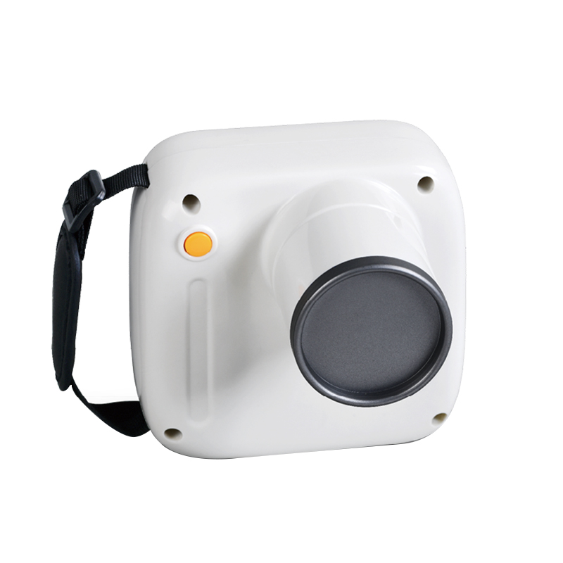
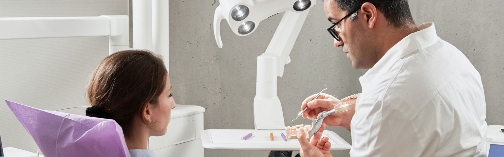
UMG manufactures intraoral dental X-ray equipment in Tangshan, China: handheld and chair-side units that expose the film or sensor, and the digital sensors that capture the image. Fifteen products sit in the range, and they are not variations of one machine — they answer three different questions, which is why this page is organised by the way the equipment is mounted and used rather than by the type of radiograph a dentist can take.
Most clinics that reach this page already know they need an intraoral X-ray. What they are deciding is whether to add a portable unit that can move between rooms, a chair-side unit with a touch control panel, or a sensor that turns an existing X-ray head into a digital workflow. Each route has a different cost, a different installation and a different set of accessories, and they are set out below in that order.
THREE ROUTES INTO INTRAORAL IMAGING
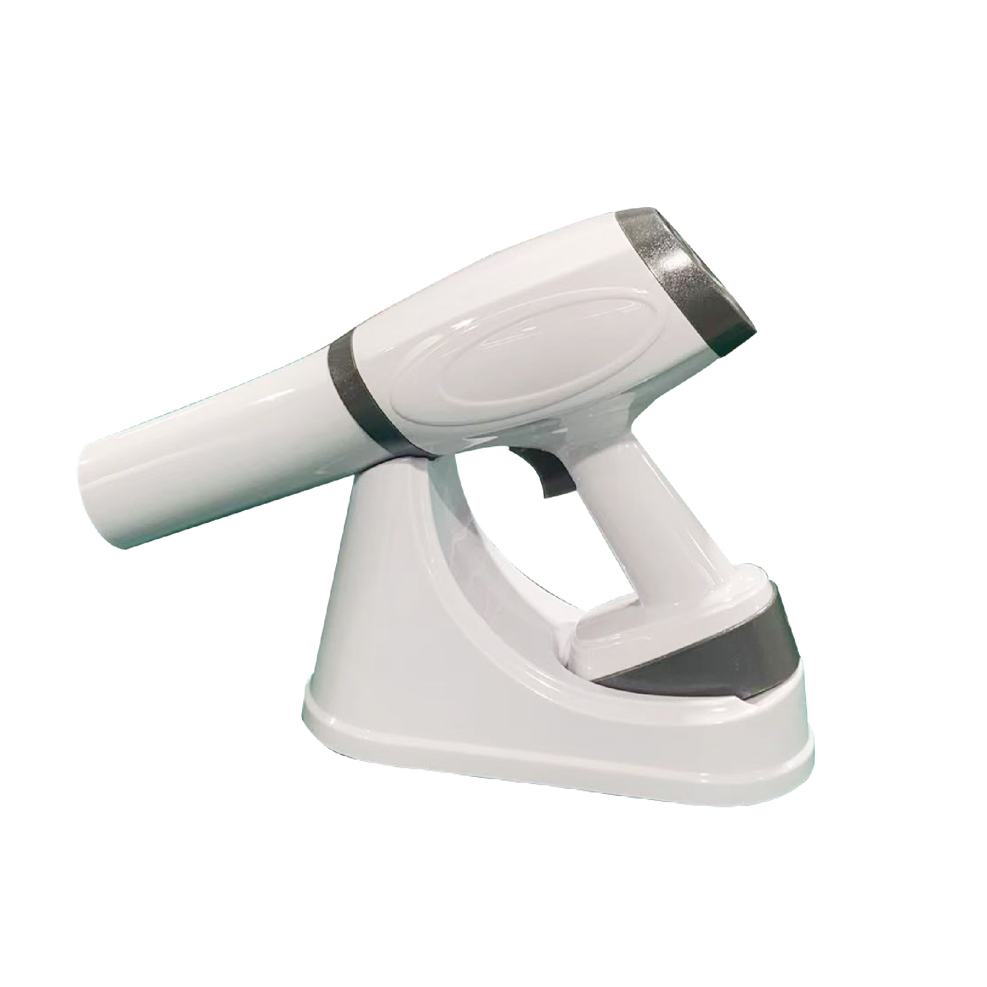 ROUTE 01 Portable unitBattery-powered, carried to the chair, no installation and no control room. |
 ROUTE 02 Chair-side unitFixed to the treatment room, with a touch panel that sets exposure time in seconds. |
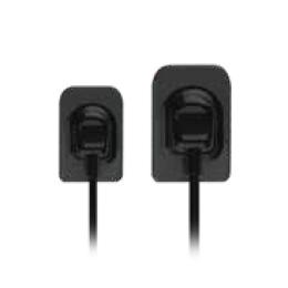 ROUTE 03 Digital sensorKeeps the X-ray head you own and replaces film with a sensor and a USB link to the PC. |
Route 01 covers eight models: UM-680, the GH series in three body shapes (standards, gun and square), UM-1S, UM-10S, X-12D and BLX-5. Route 02 is the UM-120RC. Route 03 is the sensor families — UM2121/UM2530, UMG-YR0001/YR0002 and HDR500/HDR600 — which are usually bought by a clinic that already has a working intraoral unit and wants to stop processing film.
What this page does not cover is film processing, the type of radiograph a clinician chooses, or the maintenance of an existing unit — those belong to the clinical literature and to the product manuals. What it does cover is the equipment decision, because the three routes above are not steps in a sequence: a clinic picks one, and the rest of the purchase follows from it.
Clinics fitting out a treatment room tend to buy the X-ray together with the dental chair and the dental intraoral camera, because all three are mounted around the same chair and share the same treatment-room supply. Buyers comparing imaging options across the site can start from the imaging system range.
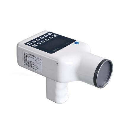
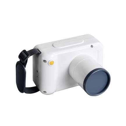
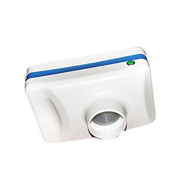
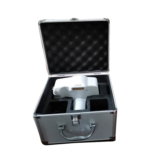
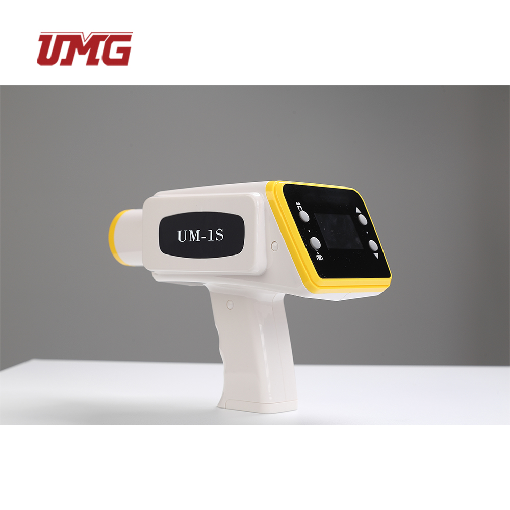
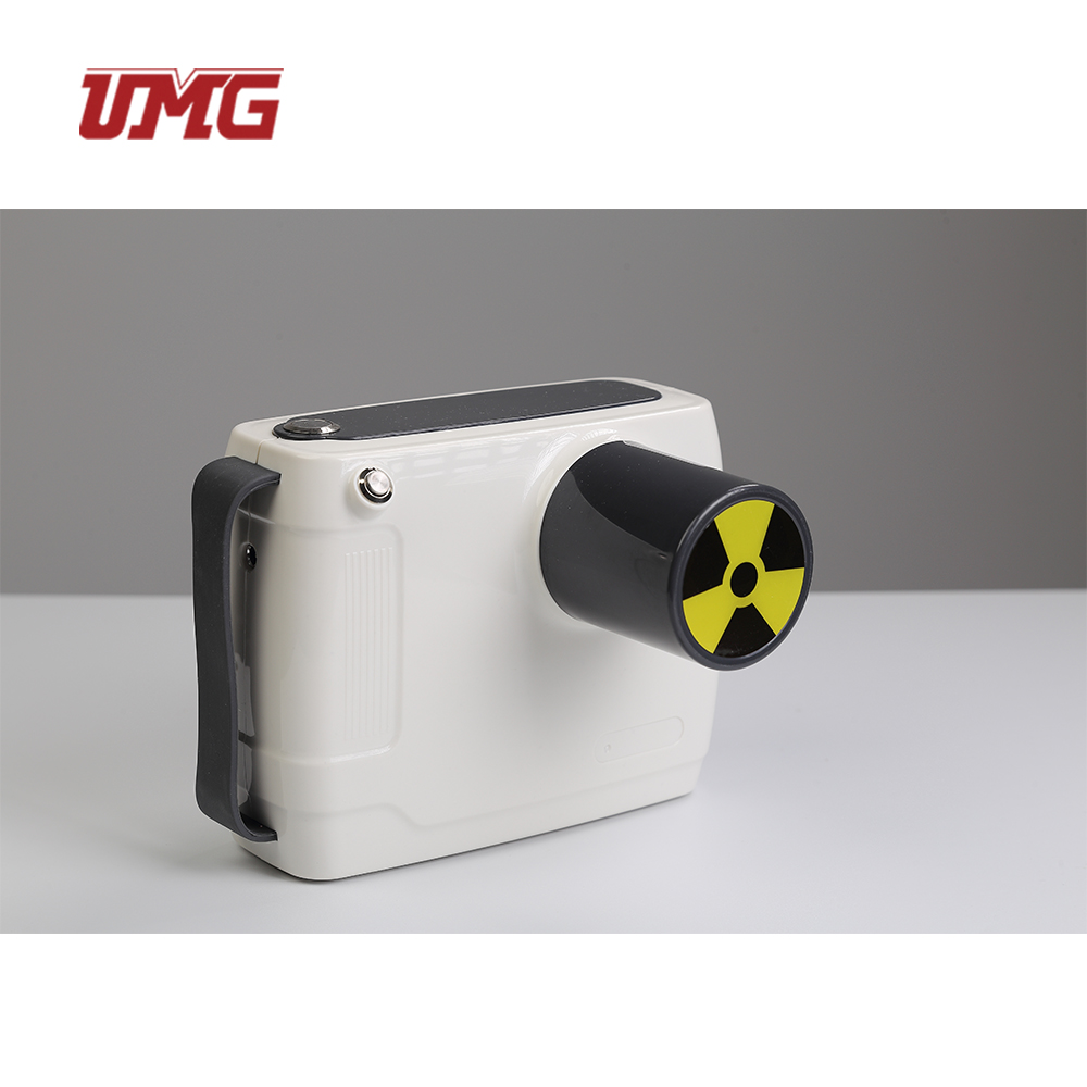
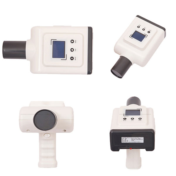
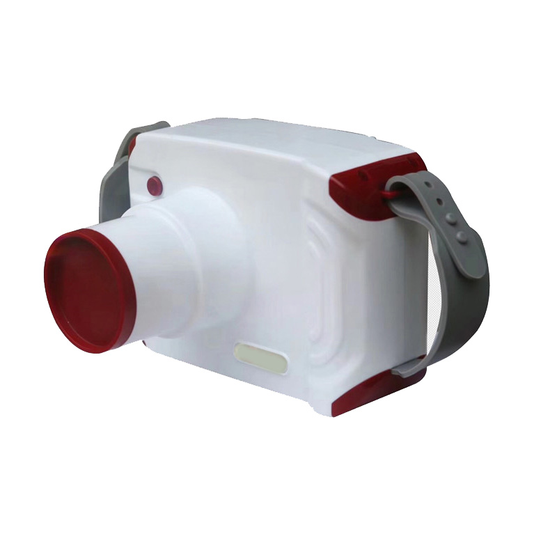
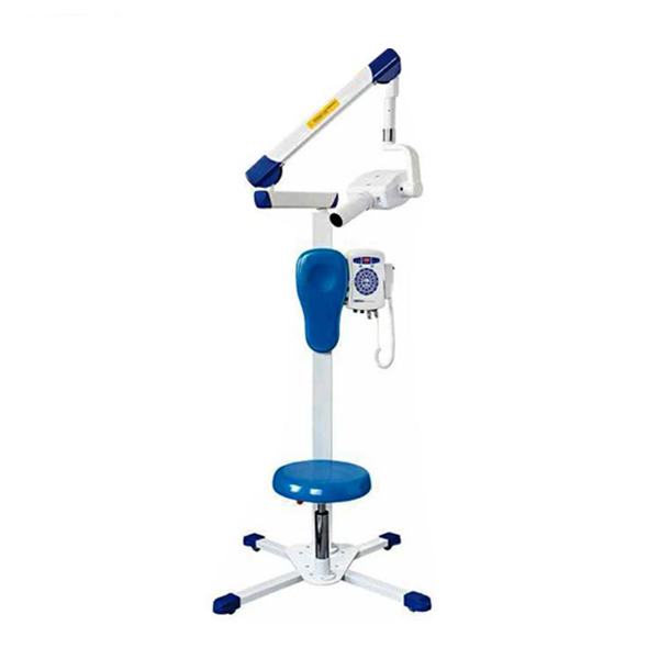
An intraoral X-ray is a small machine with a narrow job: it exposes one tooth or one group of teeth, at a short focal distance, in a fraction of a second. Everything that varies between the models below is a consequence of that job — how the tube head is held, how the exposure is timed, and whether the image arrives on film or on a screen.
SPECIFICATIONS IN COMPARISON
The four models below have published figures. All of them run a tube at 60 kV, which is the standard for intraoral work; what changes is the tube current, the exposure range and the source of power.
| Model | Form | Voltage | Current | Exposure | Power source |
| UM-680 | Handheld | 60 kV | 2 mA | 0.1–2.0 s | 14.8 V / 2,500 mAh |
| X-12D | Handheld, 1.8 kg | 60 kV | 0.28 mA | 0.3–1.5 s | 24 V / 5,000 mAh |
| BLX-5 | Portable, mains | 60 kV | 0.1 mA | 1–9 s | AC 220 V, 60 W |
| UM-120RC | Chair-side, touch screen | 60 kV | 2 mA | 0.01–2 s | Inverter above 100 kHz |
The GH series (standards, gun and square body), UM-1S and UM-10S are quoted on request with their own figures. On the UM-120RC the ripple ratio is quoted below 1% and the exposure is set on the touch panel in hundredths of a second, which matters when a paediatric exposure is being shortened rather than a molar exposure lengthened.
A handheld unit and a chair-side unit are not competing for the same buyer. The handheld machine is chosen by a clinic that has to move between treatment rooms, by a surgeon working in a second location, or by a practice that cannot run a fixed supply to the chair. The chair-side machine is chosen where the room is fixed and the exposure is set at the panel rather than at the cone — and where a higher tube current shortens the exposure.
SENSORS AND RETROFIT
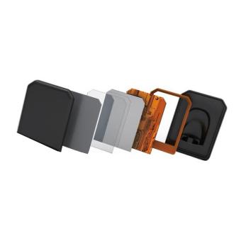
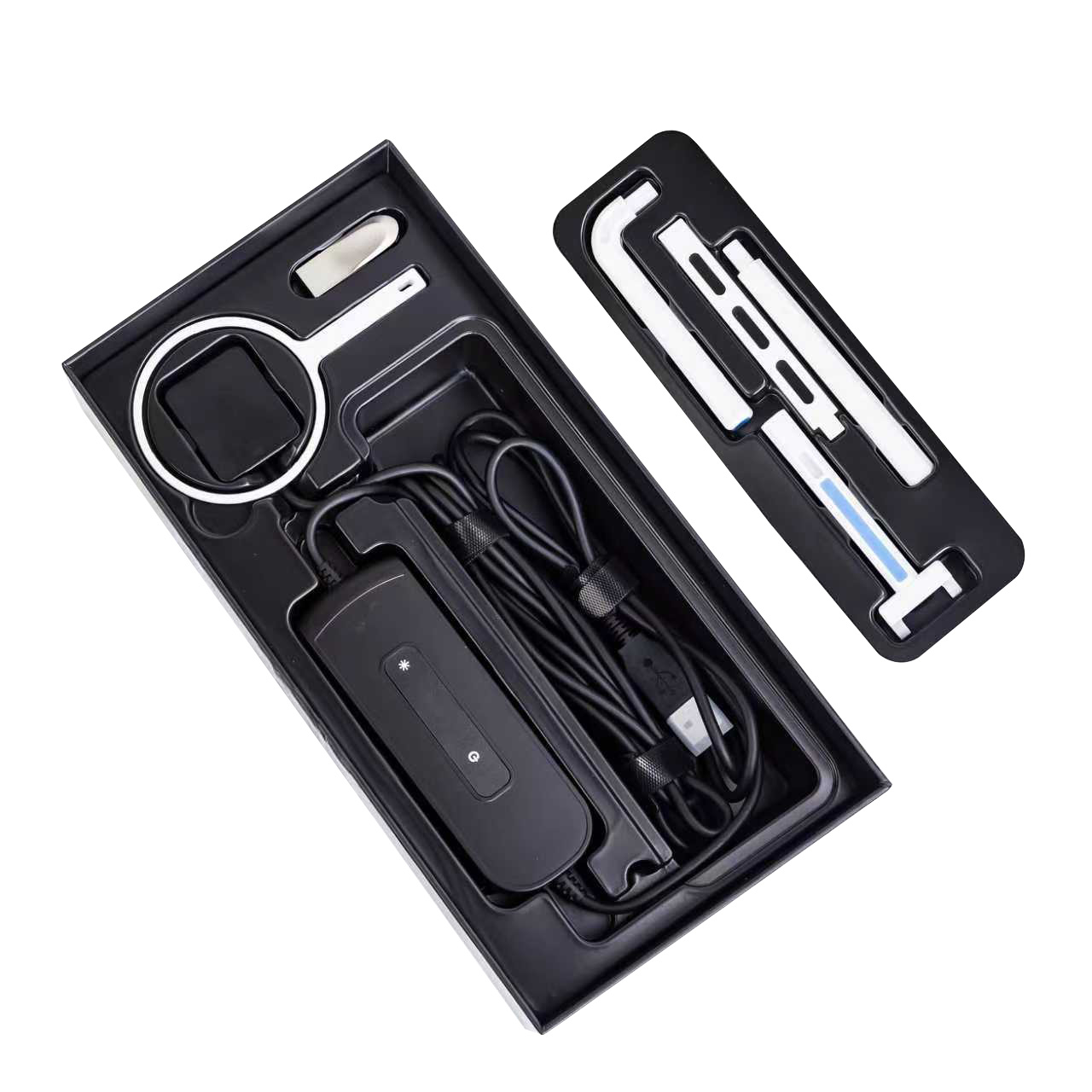
A digital sensor replaces the film packet and the processing that follows it. The sensor sits in the mouth in the same position the film would take, the existing tube head exposes it, and the image is transferred by USB to the PC at the chair. Three sensor families are stocked: UM2121/UM2530, UMG-YR0001/YR0002 and HDR500/HDR600.
This is the route with the smallest change to the practice. Nothing in the treatment room is rebuilt, no control room is added, and the exposure settings on the existing unit stay where they are — what changes is what happens after the exposure. It is also the route that most often decides whether a clinic keeps a working film unit instead of replacing it, because the head is usually the part that is still healthy.
The sensor and the unit have to be matched: the active area has to cover the teeth being imaged, the cable has to reach the PC without crossing the working field, and the imaging software has to accept the driver. Send us the X-ray unit you are keeping and the PC operating system, and we will confirm the sensor family before the order rather than after.
PERIAPICAL WORK
A periapical exposure covers the whole length of a tooth including the apex, and it is the exposure most intraoral units in this range are bought for — root canal work, apical pathology, a single implant site. Two figures decide how easily a unit does it. The first is the field: on the UM-680 the line-field limiting diameter is 58 mm, which frames a periapical view without the cone having to be repositioned between the mesial and distal surfaces. The second is the focal distance: 100 mm from focal spot to skin on the handheld models, which is the distance the cone has to be held at, and the reason a handheld unit is held with a steady grip rather than braced against the wall.
Focal spot size is the third number worth comparing, and it is 1.5 mm on the X-12D. A smaller focal spot sharpens the image at the same exposure; a larger spot tolerates a shorter exposure in a restless patient, so neither is simply better. Ask for this figure on any model you compare, because it is not always printed on the sheet.
Wholesale buyers ordering for a distributor network usually take the periapical units in mixed cartons with sensors, because a clinic buying one usually buys the other. Mixed orders are quoted as one shipment.
BEFORE THE ORDER
QUESTIONS WE ARE ASKED
Buy the portable if the unit has to move — between rooms, between floors, or to a second site — or if the room cannot take a fixed installation. Buy the chair-side unit if the room is permanent and the exposure is set at the chair. The portable saves installation; the chair-side saves handling time in a busy room.
Usually yes, provided the unit's exposure can be set low enough for a sensor and the sensor's active area suits the teeth being imaged. Send the make and model of the existing unit, the exposure range and the PC operating system, and we will confirm the sensor family that fits.
The units will expose film, and the BLX-5 is specified for daylight film processing, so no darkroom is needed. Most clinics now pair the unit with a sensor instead, which removes the processing step entirely.
All the intraoral units on this page take periapical exposures; what differs is the field size and the focal distance. The UM-680's 58 mm field and 100 mm focal distance are the figures to compare against the way your clinicians position the cone.
Yes. Units and sensors are quoted for single-clinic orders and for distributor orders, and a mixed order of units, sensors and mounting accessories ships as one consignment. Tell us the market and the volumes and we will quote accordingly.
Send the exposures you need to take, the number of rooms and whether you are keeping an existing X-ray head, and we will recommend the route and the model. Request a quotation for dental X-ray equipment.